A Well Labelled Diagram Of The Pelvic Girdle

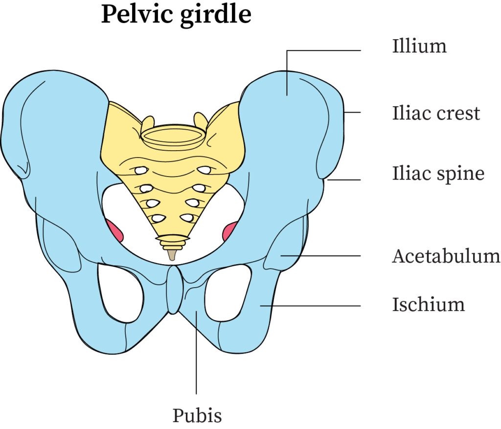
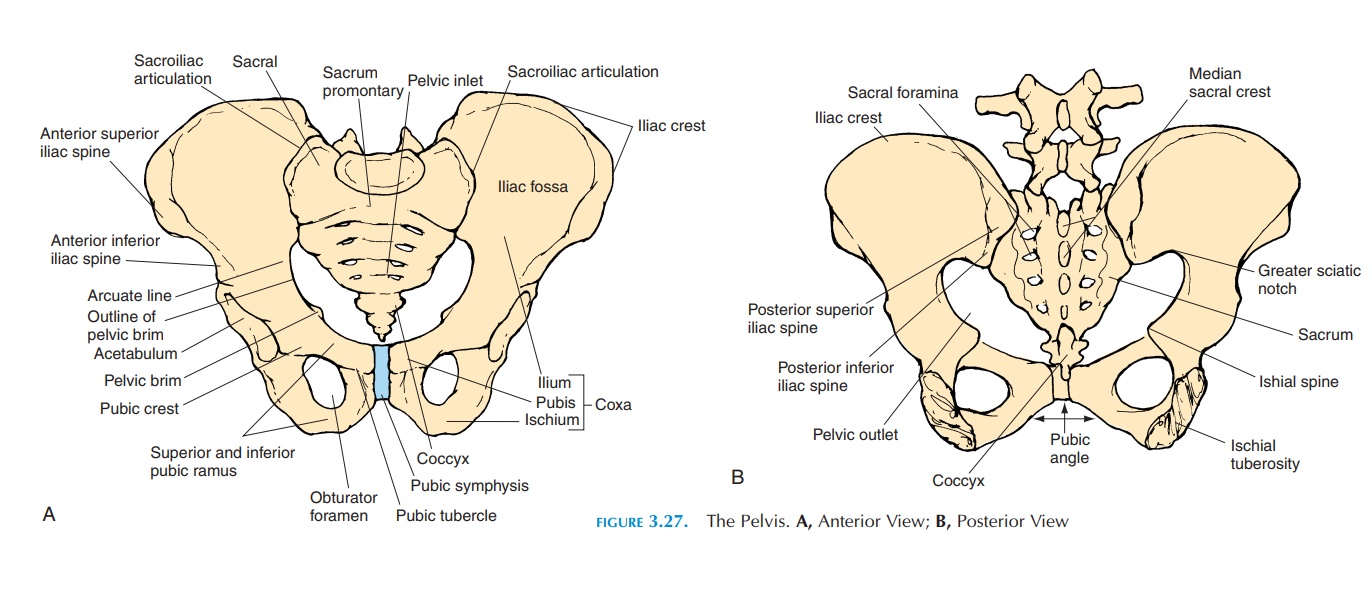
A Well Labelled Diagram Of The Pelvic Girdle Structure of the pelvic girdle. the bony pelvis consists of the two hip bones (also known as innominate or pelvic bones), the sacrum and the coccyx. there are four articulations within the pelvis: sacroiliac joints (x2) – between the ilium of the hip bones, and the sacrum. sacrococcygeal symphysis – between the sacrum and the coccyx. Figure 8.3.1 – pelvis: the pelvic girdle is formed by a single hip bone. the hip bone attaches the lower limb to the axial skeleton through its articulation with the sacrum. the right and left hip bones, plus the sacrum and the coccyx, together form the pelvis. hip bone. the hip (or coxal) bones form the pelvic girdle portion of the pelvis.

A Well Labelled Diagram Of The Pelvic Girdle The pelvic girdle (hip girdle) is formed by a single bone, the hip bone or coxal bone (coxal = “hip”), which serves as the attachment point for each lower limb. each hip bone, in turn, is firmly joined to the axial skeleton via its attachment to the sacrum of the vertebral column. the right and left hip bones also converge anteriorly to. Alex aranda, elizabeth nixon shapiro, msmi, cmi, ursula florjanczyk, mscbmc. the pelvic girdle or the bony pelvis is a bony ring, formed by the left and right hip bones and the sacrum, and it surrounds the pelvic cavity, and connects the vertebral column to the lower limbs. the main functions of the pelvic girdle are to transfer the weight of. Anatomy of the pelvic girdle. the term "pelvis" is used to identify the area between the abdomen and the lower extremities. it can be divided into the greater pelvis and the lesser pelvis. [1] the pelvis consists of the sacrum, the coccyx, the ischium, the ilium, and the pubis. [2] [3] the structure of the pelvis supports the contents of the. The pelvic floor is formed by the bowl or funnel shaped pelvic diaphragm, consisting of the levator ani and coccygeus muscles and their investing fascia. structurally, the pelvic floor separates the pelvic cavity from the perineum. functionally, these pelvic floor muscles support the pelvic organs, keeping them in place and preventing prolapse.

Labeled Diagram Of Pelvic Girdle Anatomy of the pelvic girdle. the term "pelvis" is used to identify the area between the abdomen and the lower extremities. it can be divided into the greater pelvis and the lesser pelvis. [1] the pelvis consists of the sacrum, the coccyx, the ischium, the ilium, and the pubis. [2] [3] the structure of the pelvis supports the contents of the. The pelvic floor is formed by the bowl or funnel shaped pelvic diaphragm, consisting of the levator ani and coccygeus muscles and their investing fascia. structurally, the pelvic floor separates the pelvic cavity from the perineum. functionally, these pelvic floor muscles support the pelvic organs, keeping them in place and preventing prolapse. Pelvic girdle. four images are shown; a photo and diagram of the anterosuperior view of the pelvic girdle, and a photo and diagram of the right hip bone. the first image has labeled parts bony pelvis, coxal bones, pubic symphysis, sacroiliac joint, sacrum, and coccyx. in the second image, the pelvic girdle has the coxal bone on the side. The pelvic girdle is part of the appendicular skeleton, which also includes the shoulder girdle and the upper and lower limbs. the ilium is the largest and most recognizable part of the pelvis: it looks like the top of a wing. if your hip bones "stick out" (are visible through your skin), it's usually the ilium you're seeing; they protrude.

A Well Labelled Diagram Of The Pelvic Girdle Pelvic girdle. four images are shown; a photo and diagram of the anterosuperior view of the pelvic girdle, and a photo and diagram of the right hip bone. the first image has labeled parts bony pelvis, coxal bones, pubic symphysis, sacroiliac joint, sacrum, and coccyx. in the second image, the pelvic girdle has the coxal bone on the side. The pelvic girdle is part of the appendicular skeleton, which also includes the shoulder girdle and the upper and lower limbs. the ilium is the largest and most recognizable part of the pelvis: it looks like the top of a wing. if your hip bones "stick out" (are visible through your skin), it's usually the ilium you're seeing; they protrude.

Comments are closed.