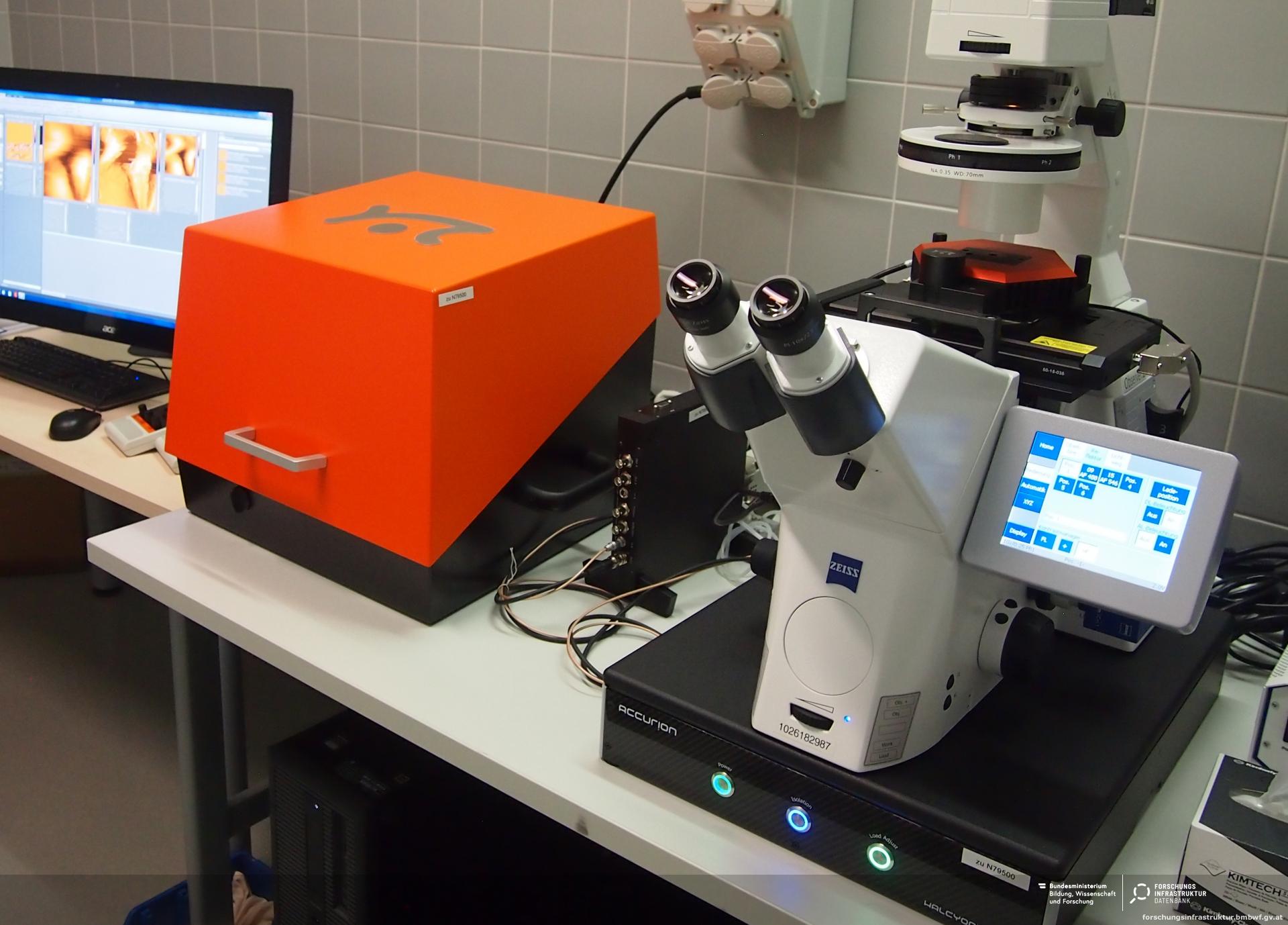
Afm Atomic Force Microscope University Of Greifswald

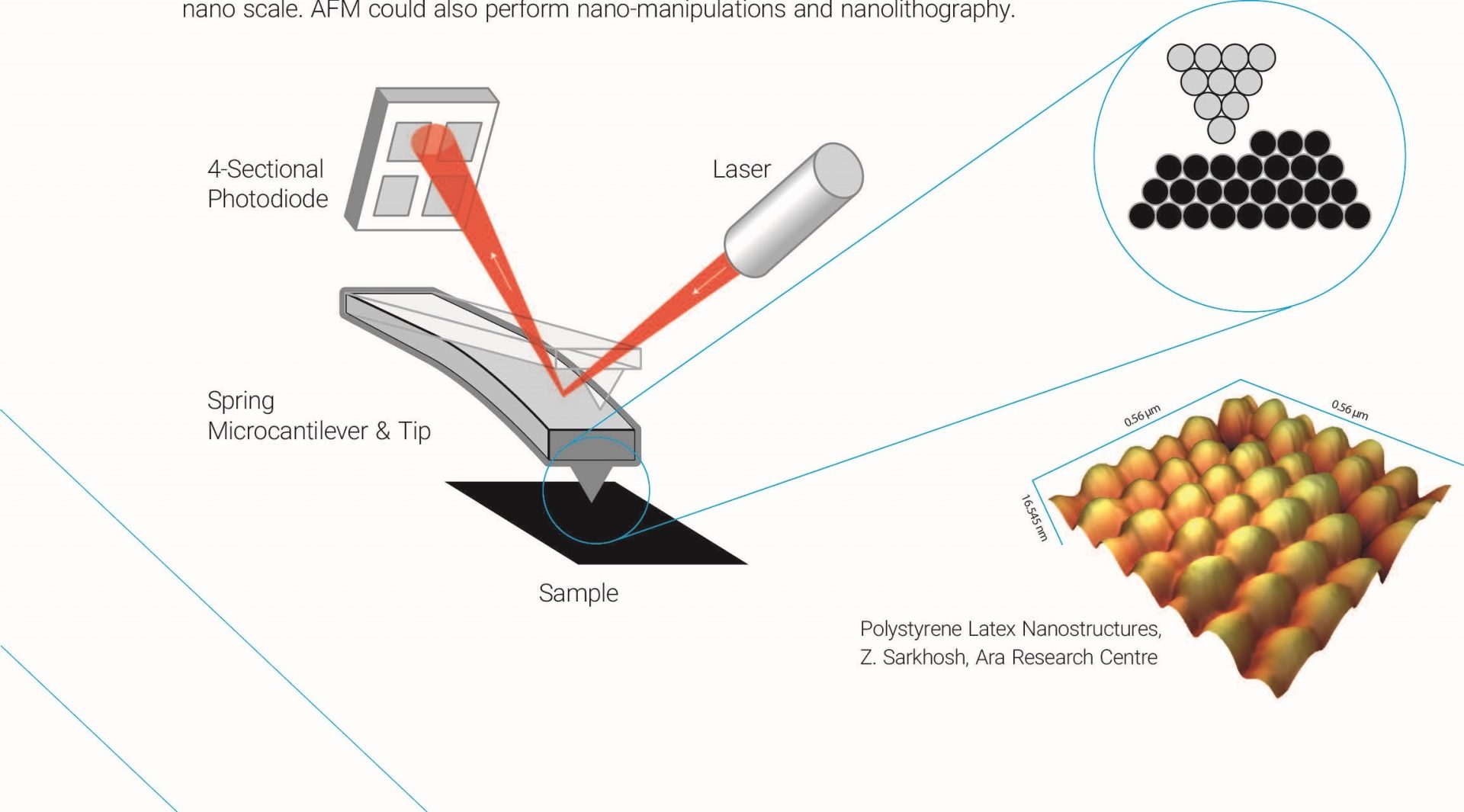
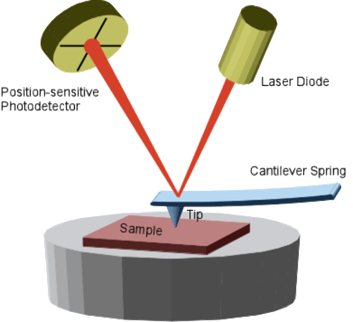
Atomic Force Microscope Afm Forschungsinfrastruktur Principles. sem picture of tip the atomic force microscope (afm) is one type of scanning probe microscopes, which is used to image surface structures (on a nm or even sub nm scale scale) and to measure surface forces. the standard afm contains a microscopic tip (curvature radius of ~10 50nm) attached to a cantilever spring. In fact, more than 10 years have passed since the discovery of nets, and although their importance as part of a unique cellular response mechanism has become very clear, studies that attempt to address them by atomic force microscopy (afm) remain very limited.

Atomic Force Microscope Spm Complete System Srl In this work, using a combination of fluorescence and atomic force microscopy, we show that nets appear as a branching filament network that results in a substantially organized porous structure with openings with 0.03 ± 0.04 μm(2) on average and thus in the size range of small pathogens. Metrics. atomic force microscopy (afm) is unique in visualizing functional biomolecules in aqueous solution at ~1 nm resolution. by borrowing localization methods from fluorescence microscopy, afm. Here, nanoindentation based atomic force microscopy (afm) was used to measure the elasticity of human embryonic kidney cells in the presence and absence of these pharmaceuticals. the results showed that depletion of cholesterol in the plasma membrane with mβcd resulted in cell stiffening whereas depolymerization of the actin cytoskeleton by. High speed atomic force microscopy (hs afm) is a powerful technique that enables real space and real time observations of macromolecules, which are not feasible by other techniques 19.hs afm.

Afm Atomic Force Microscope University Of Greifswald Here, nanoindentation based atomic force microscopy (afm) was used to measure the elasticity of human embryonic kidney cells in the presence and absence of these pharmaceuticals. the results showed that depletion of cholesterol in the plasma membrane with mβcd resulted in cell stiffening whereas depolymerization of the actin cytoskeleton by. High speed atomic force microscopy (hs afm) is a powerful technique that enables real space and real time observations of macromolecules, which are not feasible by other techniques 19.hs afm. The scanning tunneling microscope (stm) and the atomic force microscope (afm) are scanning probe microscopes capable of resolving surface detail down to the atomic level. the potential of these microscopes for revealing subtle details of structure is illustrated by atomic resolution images including graphite, an organic conductor, an insulating. The ability to probe a material’s electromechanical functionality on the nanoscale is critical to applications from energy storage and computing to biology and medicine. voltage modulated atomic force microscopy (vm afm) has become a mainstay characterization tool for investigating these materials due to its ability to locally probe electromechanically responsive materials with spatial.

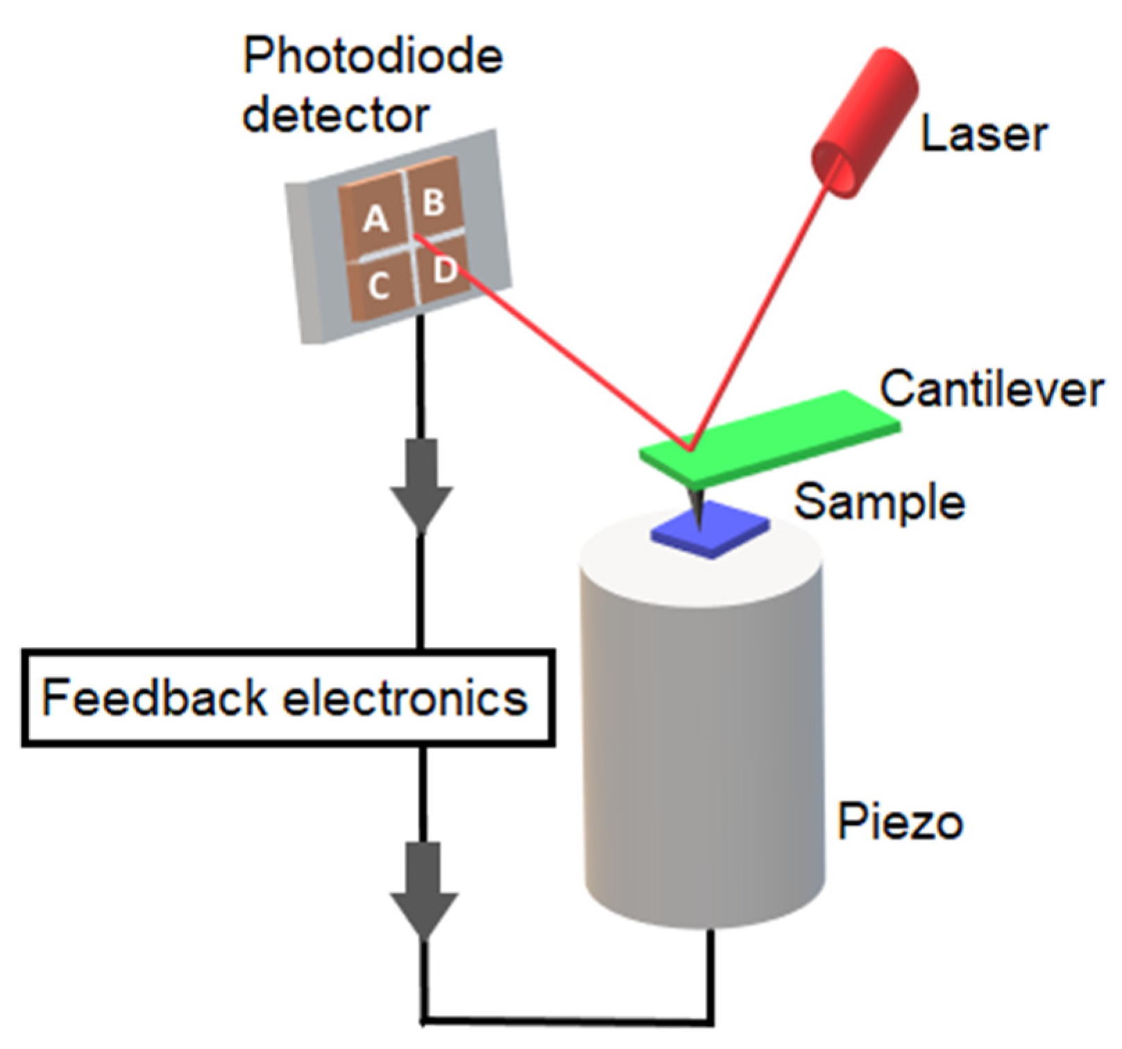
Water Free Full Text Insights Into The Morphology And Surface The scanning tunneling microscope (stm) and the atomic force microscope (afm) are scanning probe microscopes capable of resolving surface detail down to the atomic level. the potential of these microscopes for revealing subtle details of structure is illustrated by atomic resolution images including graphite, an organic conductor, an insulating. The ability to probe a material’s electromechanical functionality on the nanoscale is critical to applications from energy storage and computing to biology and medicine. voltage modulated atomic force microscopy (vm afm) has become a mainstay characterization tool for investigating these materials due to its ability to locally probe electromechanically responsive materials with spatial.

Imaging

Comments are closed.