Anatomy Pathology And Radiography Ppt Download

Ppt Radiographic Anatomy And Positioning Of The Upper Extremity Anatomy cranium the ***skull consists of two parts – the cranium and the facial bones. the cranium is divided into two sections, the floor and the calvaria. the floor of the cranium is made up of the ethmoid, sphenoid, and 2 temporal bones. the calvaria is made of the frontal, occipital, and 2 parietals. ***orbits are commonly referred to as eye sockets. each orbit is composed of 7 bones 3. 16 ribs radiography to image anterior ribs, position front close to ir. to image posterior, put the patient’s back to the ir. imaging the ribs is done with the side closest to the ir. so, if the anterior ribs are suspected of having a pathology, the patient would be placed in the pa position with the front of his ribs close to the ir.

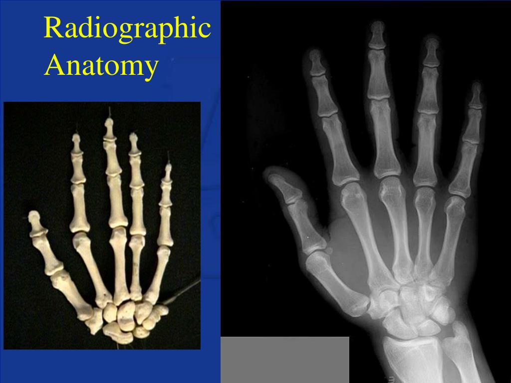
Ppt Normal Radiographic Anatomy Powerpoint Presentation Free 1. the document contains radiographic images and descriptions of normal anatomy across multiple body regions. images include chest, abdomen, spine, shoulder, elbow, wrist, hand, pelvis, hip, knee, ankle, foot, skull and cervical spine. 2. key normal structures are labeled on the images, such as bones, joints, organs and vasculature. The most frequently used imaging modalities are radiography (x ray), computed tomography (ct) and magnetic resonance imaging (mri). x ray and ct require the use of ionizing radiation while mri uses a magnetic field to detect body protons. mri is the safest among the three, although each technique has its benefits. This document provides an overview of pelvic anatomy and normal pelvic radiology. it describes the bones of the pelvis, ligaments, muscles, blood vessels and lymph nodes. examples of normal anatomy are shown on plain radiographs, ct scans and mri images in axial, sagittal and coronal views. key structures like the sacrum, hip bones, bladder and. Aug 06, 2014. 460 likes | 965 views. abdomen radiography. abdomen module . sean duguay and justin north, md. directions. view in presentation mode this module is meant for independent study highlighted words in yellow have actions clicking on them once will identify certain areas. download presentation.

Comments are closed.