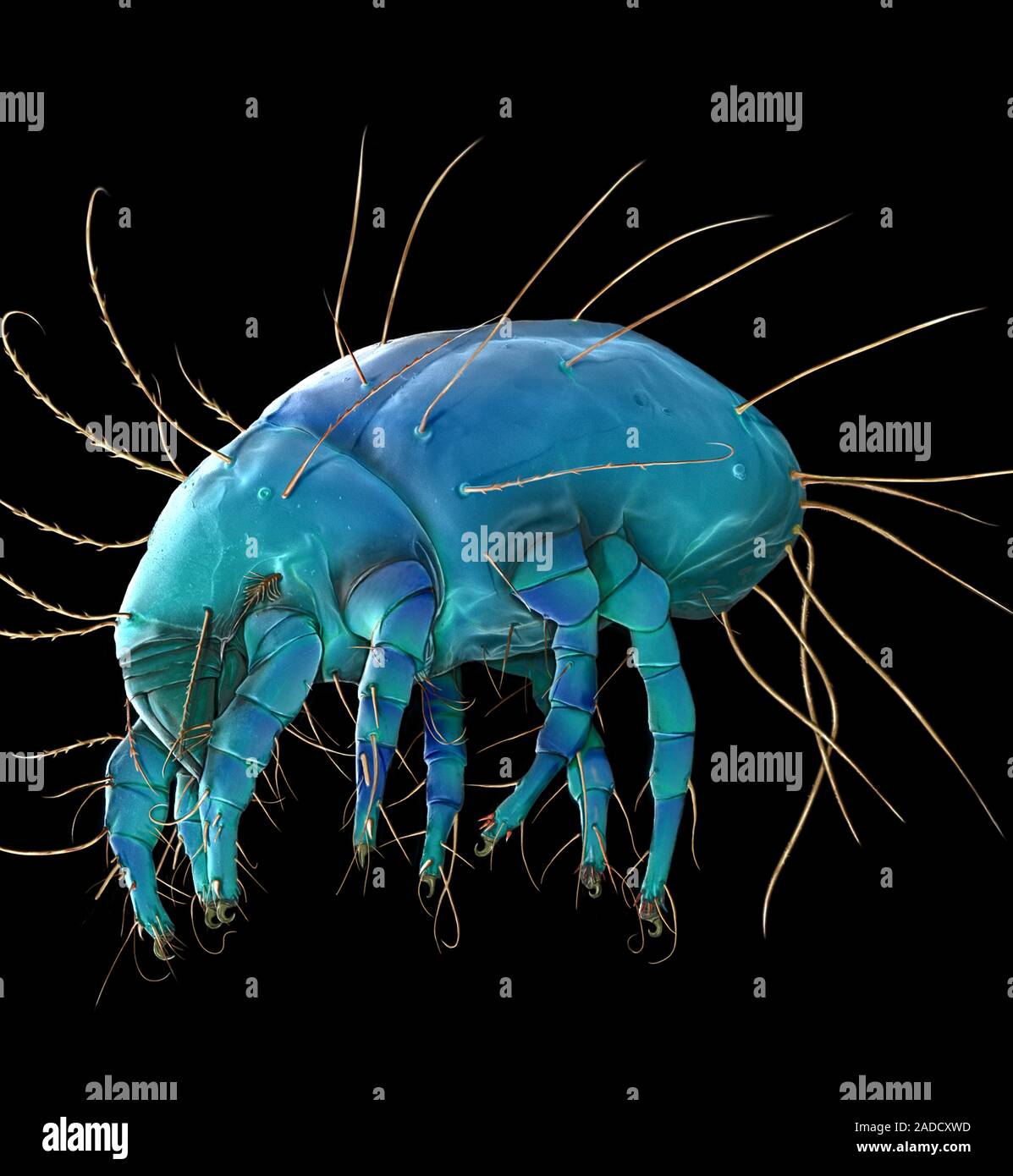
Coloured Scanning Electron Micrograph Sem Of Dust Mite

Coloured Scanning Electron Micrograph Sem Of Photocomposite Of Dust Coloured scanning electron micrograph (sem) of dust mite (dermatophagoides pteronyssinus). millions of dust mites inhabit the home, feeding on dead human skin that are common in house dust. the mite's body is in three parts: the gnathosoma (head region) adapted for feeding on dead skin, the propodosma (carrying the 1st and 2nd pair of walking. Dust mite. coloured scanning electron micrograph (sem) of a house dust mite (dermatophagoides pteronyssinus). millions of dust mites inhabit the home, feeding on shed skin cells. they mainly live in furniture, and are usually harmless. however, their excrement and dead bodies may cause allergic reactions in susceptible people.

Dust Mite Coloured Scanning Electron Micrograph Sem Of A H Scanning electron microscope (sem) images of dust mites, suidasia pontifica, is presented to provide an improved visualization of the taxonomic characters of these mites. suidasia pontifica can easily be identified by its scale like cuticle, presence of external vertical setae (ve), longer external scapular setae (sce) compared to internal. Download this stock image: coloured scanning electron micrograph (sem) of dust mite (dermatophagoides pteronyssinus).millions of dust mites inhabit home,feeding on dead human skin that are common in house dust.the mite's body is in three parts: gnathosoma (head region) adapted for feeding on dead skin,the propodosma hj0c1c from alamy's library of millions of high resolution stock photos. 2 electron microscopy unit, institute for medical research, 50588 kuala lumpur * corresponding author email: [email protected] received 8 september 2010; received in revised form 14 january 2011; accepted 20 january 2011 abstract. scanning electron microscope (sem) images of dust mites, suidasia pontifica, is. Scanning electron microscope (sem) images of two dust mites, sturnophagoides brasiliensis and sturnophagoides halterophilus, are presented to provide an improved visualization of the taxonomic.

Comments are closed.