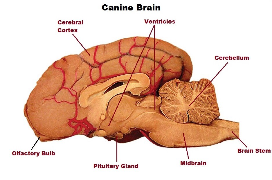
Dog Brain Anatomy

Dog Brain Anatomy Explore the anatomy of the dog brain with 12 transverse levels of images and a glossary. learn about the canine brain transections, mri atlas and veterinary anatomy web site. Explore transverse views of a beagle brain obtained by magnetic resonance imaging and paired with stained tissue sections. learn brain anatomy with interactive quizzes and mri image interpretation tips.

Canine Brain Anatomy Movement Science Motor Control At Queen Mary Exit to veterinary anatomy web site. canine brain transections is a website intended for veterinary students studying neuroanatomy. it presents neuroanatomy of the canine brain as seen in transverse sections. this website has two divisions, which students may toggle between per brain transection level:. The brain is divided into 3 main sections—the brain stem, which controls many basic life functions, the cerebrum, which is the center of conscious decision making, and the cerebellum, which is involved in movement and motor control. the spinal cord of dogs is divided into regions that correspond to the vertebral bodies (the bones that make up. Explore the normal anatomy of the dog brain on mri with more than 400 images and 305 labeled parts. learn the latin and english terms for the cerebral lobes, ventricles, arteries, nerves and more. A population based average of 15 mesaticephalic dog brains derived from in vivo and ex vivo mri data. the atlas includes a brain template, a high resolution template, and a surface representation of the gray matter white matter boundary.

1 Canine Brain Dr Bills Pet Nutrition Explore the normal anatomy of the dog brain on mri with more than 400 images and 305 labeled parts. learn the latin and english terms for the cerebral lobes, ventricles, arteries, nerves and more. A population based average of 15 mesaticephalic dog brains derived from in vivo and ex vivo mri data. the atlas includes a brain template, a high resolution template, and a surface representation of the gray matter white matter boundary. Several brain atlases have been made available for the canine 10,11,12, however these atlases have limitations, being created from a low number of subjects 10, using non isovolumetric clinical. Here we present a canine brain atlas derived as the diffeomorphic average of a population of. fifteen mesaticephalic dogs. the atlas includes: 1) a brain template derived from in vivo, t1 weighted.

Comments are closed.