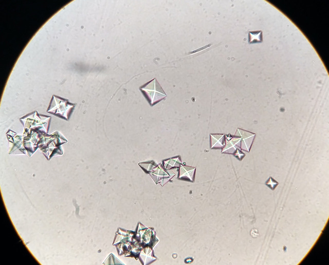
Figure Microscopic View Of Calcium Oxalate Statpearls Ncbi

Figure Microscopic View Of Calcium Oxalate Statpearls Ncbi Ncbi bookshelf. a service of the national library of medicine, national institutes of health. microscopic view of calcium oxalate crystals in urine. created by. Urinary crystal formation is a marker of urinary supersaturation with various substances. crystals can occur secondary to inherited diseases, metabolic disorders, and medications. the presence of crystals in the urine is not always pathognomic for an abnormal metabolic or renal condition, as uric acid, calcium oxalate, calcium phosphate (cap), and drug induced crystals can also be observed.

Figure These Pictures Display The Calcium Statpearls Ncbi Renal calculi are the products of crystallization of specific stone forming components seen in about 10% of people, and 80% are calcium based stones. the most common stone, calcium oxalate, is formed primarily due to an imbalance between calcium and oxalate levels in the body or a lack of adequate crystallization inhibitors. hyperoxaluria is a significant contributor to nephrolithiasis, and. Calcium oxalate stones are the most common type of renal calculi, comprising 70% to 75% of all urinary stones. while chemically identical, they may present as 2 different crystalline forms: calcium oxalate monohydrate (whewellite, very hard) or a dihydrate (weddelite, brittle). these stones typically form in acidic urine but may be found with. The kidneys are a primary site of crystalline deposition, receiving 25% of total cardiac output. these deposits primarily occur in the renal tubules, interstitium, or both. they are associated with the development of acute kidney injury (aki) or progressive chronic kidney disease (ckd), which can present with microscopic hematuria, crystalluria. The most common stone, calcium oxalate, is formed primarily due to an imbalance between calcium and oxalate levels in the body or a lack of adequate crystallization inhibitors. hyperoxaluria is a significant contributor to nephrolithiasis, and the causes of excess urinary oxalate can be classified into primary and secondary hyperoxaluria based.

The Calcium Oxalate Crystals Viewed Under Light Microscope 400x In The kidneys are a primary site of crystalline deposition, receiving 25% of total cardiac output. these deposits primarily occur in the renal tubules, interstitium, or both. they are associated with the development of acute kidney injury (aki) or progressive chronic kidney disease (ckd), which can present with microscopic hematuria, crystalluria. The most common stone, calcium oxalate, is formed primarily due to an imbalance between calcium and oxalate levels in the body or a lack of adequate crystallization inhibitors. hyperoxaluria is a significant contributor to nephrolithiasis, and the causes of excess urinary oxalate can be classified into primary and secondary hyperoxaluria based. Hypercalciuria is generally considered to be the most common identifiable metabolic risk factor for calcium nephrolithiasis. it also contributes to osteopenia and osteoporosis. its significance is primarily due to these two clinical entities: nephrolithiasis and bone resorption. calcium based kidney stones (calcium oxalate and calcium phosphate. Nephrocalcinosis, a term first coined in 1934 by fuller albright, refers to augmented calcium content within the kidney which can be in the form of calcium oxalate or calcium phosphate. however, there have been suggestions that the definition of nephrocalcinosis should be limited to calcium phosphat ….

Comments are closed.