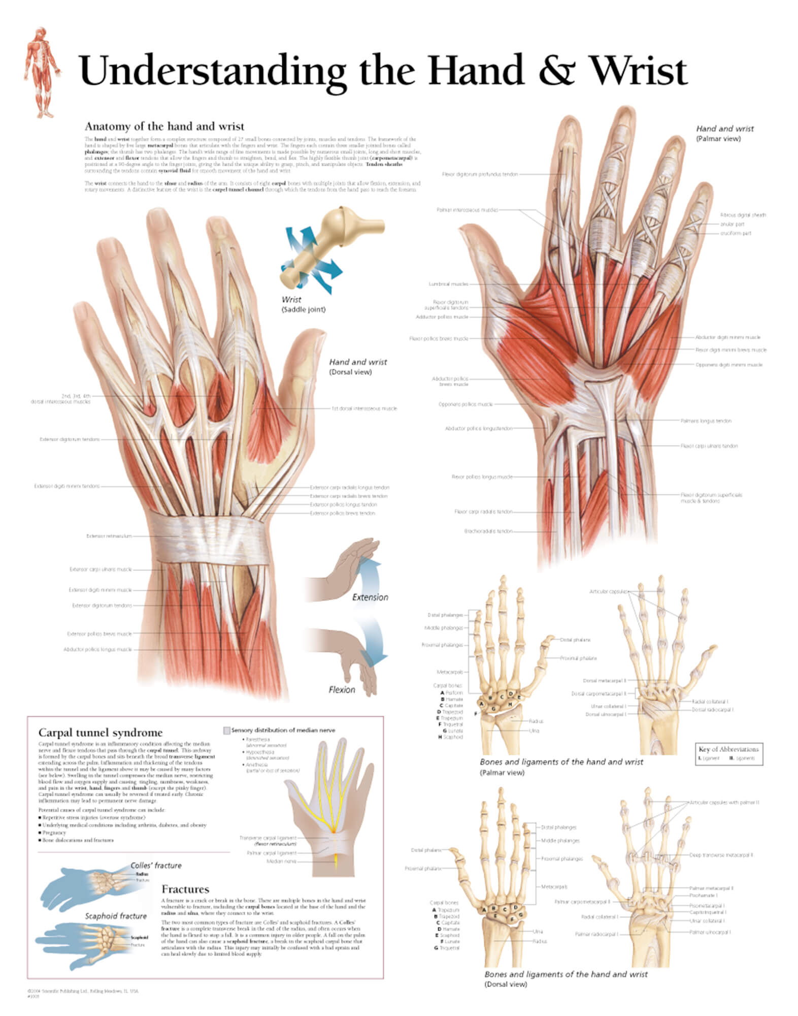
Hand And Wrist Anatomical Chart

Understanding The Hand Wrist Scientific Publishing Learn about the bones, muscles, nerves, tendons and ligaments that make up your hand and wrist. see diagrams and descriptions of the different parts and functions of your hand and wrist anatomy. Learn about the anatomy of the hand, including its bones, muscles, nerves, arteries and veins. find out how the hand is connected to the wrist and forearm, and explore the functions and movements of the hand muscles.

Anatomy Of The Wrist Anatomical Charts Posters Learn about the structure and function of the hand and wrist, including the 27 bones, the carpal bones, the joints, the ligaments, the muscles, the tendons, and the nerves. see anatomy pictures and diagrams of the hand and wrist anatomy. Carpal bones (proximal) – a set of eight irregularly shaped bones. they are located in the area of the wrist. metacarpals – a set of five bones, each one related to a digit. they are located in the area of the palm. phalanges (distal) – the bones of the digits. the thumb has two phalanges, whilst the rest of the fingers have three. There are 27 bones within the wrist and hand. the wrist itself contains eight small bones, called carpals. the carpals join with the two forearm bones, the radius and ulna, forming the wrist joint. further into the palm, the carpals connect to the metacarpals. there are five metacarpals forming the palm of the hand. Rotation of the radius around the ulna results in the supination and pronation of the hand. these bones also form the flexible wrist joint with the proximal row of the carpals. there are eight small carpal bones in the wrist that are firmly bound in two rows of four bones each. the mass that results from these bones is called the carpus.

Comments are closed.