Heart Anatomy Labeling

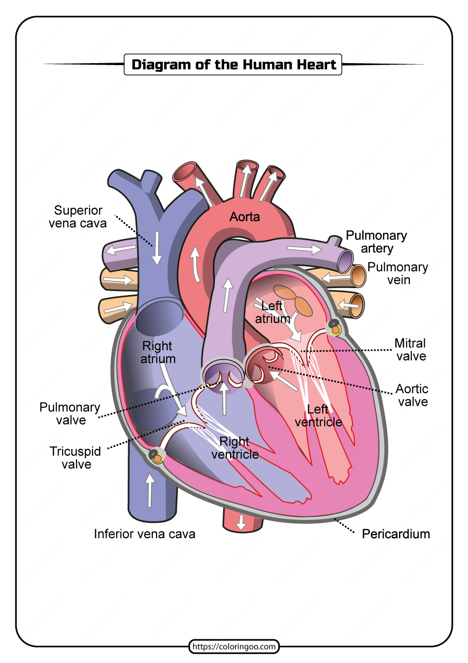
Free Printable Diagram Of The Human Heart Segment of the heart that receives deoxygenated blood. aorta. the main artery carrying oxygenated blood to all parts of the body. in this interactive, you can label parts of the human heart. drag and drop the text labels onto the boxes next to the diagram. selecting or hovering over a box will highlight each area in the diagram. Ummmmmmm . . . it's pretty self explanatory . . . you label the heart. just remember one thing you're looking at the heart like it's in someone else so right and left are switched around.

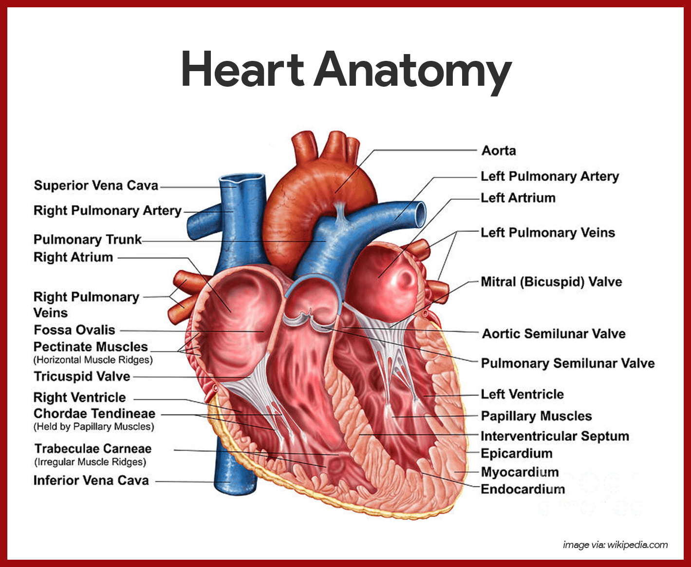
Cardiovascular System Anatomy And Physiology Study Guide For Nurses Anatomy online heart labeling quiz. the aorta is the main and largest artery in the human body. it originates from the left ventricle. the aortic valve lies at the base of the aorta. it permits blood blood to leave the left ventricle as it contracts. the inferior vena cava and superior vena cava carry blood to the right atrium. Function and anatomy of the heart made easy using labeled diagrams of cardiac structures and blood flow through the atria, ventricles, valves, aorta, pulmonary arteries veins, superior inferior vena cava, and chambers. includes an exercise, review worksheet, quiz, and model drawing of an anterior view (frontal section) of the heart in order to. The aortic semilunar valve is between the left ventricle and the opening of the aorta. it has three semilunar cusps leaflets: left left coronary, right right coronary, and posterior non coronary. in clinical practice, the heart valves can be auscultated, usually by using a stethoscope. Anatomy of the heart: anatomical illustrations and structures, 3d model and photographs of dissection. this interactive atlas of human heart anatomy is based on medical illustrations and cadaver photography. the user can show or hide the anatomical labels which provide a useful tool to create illustrations perfectly adapted for teaching.

Show Me A Diagram Of The Human Heart Here Are A Bunch The aortic semilunar valve is between the left ventricle and the opening of the aorta. it has three semilunar cusps leaflets: left left coronary, right right coronary, and posterior non coronary. in clinical practice, the heart valves can be auscultated, usually by using a stethoscope. Anatomy of the heart: anatomical illustrations and structures, 3d model and photographs of dissection. this interactive atlas of human heart anatomy is based on medical illustrations and cadaver photography. the user can show or hide the anatomical labels which provide a useful tool to create illustrations perfectly adapted for teaching. The base of the heart is located along the body's midline with the apex pointing toward the left side. because the heart points to the left, about 2 3 of the heart's mass is found on the left side of the body and the other 1 3 is on the right. anatomy of the heart pericardium. the heart sits within a fluid filled cavity called the pericardial. 1. the heart wall is composed of three layers. the muscular wall of the heart has three layers. the outermost layer is the epicardium (or visceral pericardium). the epicardium covers the heart, wraps around the roots of the great blood vessels, and adheres the heart wall to a protective sac. the middle layer is the myocardium.

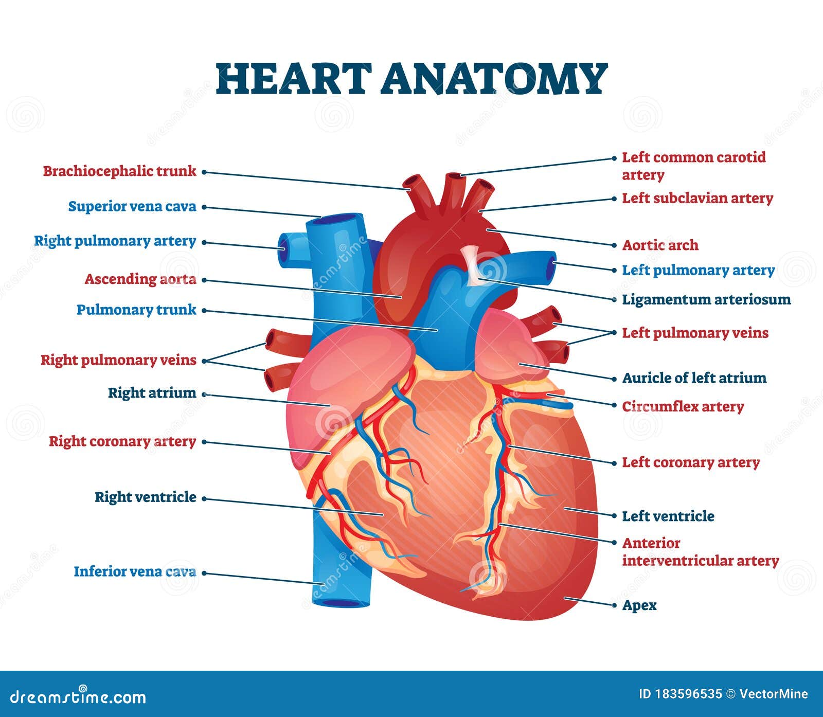
Heart Anatomy Vector Illustration Labeled Organ Structure Educational The base of the heart is located along the body's midline with the apex pointing toward the left side. because the heart points to the left, about 2 3 of the heart's mass is found on the left side of the body and the other 1 3 is on the right. anatomy of the heart pericardium. the heart sits within a fluid filled cavity called the pericardial. 1. the heart wall is composed of three layers. the muscular wall of the heart has three layers. the outermost layer is the epicardium (or visceral pericardium). the epicardium covers the heart, wraps around the roots of the great blood vessels, and adheres the heart wall to a protective sac. the middle layer is the myocardium.

Anatomy Of The Human Heart

Comments are closed.