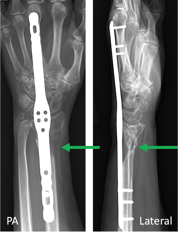
Open Reduction Internal Fixation Of Left Distal Radius Ulna

Open Reduction Internal Fixation Of Left Distal Radius Ulna The first part is open reduction. the surgeon will cut the skin and move the bone back into the normal position. the second part is internal fixation. the surgeon will attach metal rods, screws. A common form of internal fixation involves an open surgical technique in which an incision is made over the fracture and a stainless steel plate with screws is placed to align the bone ends and prevent displacement or loss of reduction. figure 8. internal fixation of a distal radius fracture. advantages of internal fixation include: increased.

Left Forearm Open Reduction And Internal Fixation Of Radius And Visit ortholibrary.org for more educational videos from nyu langone orthopedicsproduced by dylan lowe, md instagram dylanlowemd twitt. This animation depicts an open reduction and internal fixation of an open comminuted fracture and dislocation of the left distal radius and ulna. the open wo. 2 6 weeks. focus on recovery of finger, then wrist, motion within the early postoperative period. splint: fashion a removable forearm fracture brace along the full length of the forearm holding the wrist in 20 30 degrees of extension. if a cast has been applied, leave in place until week 4 and fashion the splint at that time. The two bones of the forearm (the radius and ulna) allow flexion and extension at the elbow and the wrist via diarthrodial joints. the radius and ulna exist in a delicate anatomical balance that allows for pronation and supination of the hand in a 180 degree arc of motion. this activity will briefly review the mechanism, diagnosis, and.

Open Reduction Internal Fixation And Bone Grafting Distal Radius 2 6 weeks. focus on recovery of finger, then wrist, motion within the early postoperative period. splint: fashion a removable forearm fracture brace along the full length of the forearm holding the wrist in 20 30 degrees of extension. if a cast has been applied, leave in place until week 4 and fashion the splint at that time. The two bones of the forearm (the radius and ulna) allow flexion and extension at the elbow and the wrist via diarthrodial joints. the radius and ulna exist in a delicate anatomical balance that allows for pronation and supination of the hand in a 180 degree arc of motion. this activity will briefly review the mechanism, diagnosis, and. A randomized prospective study on the treatment of intra articular distal radius fractures: open reduction and internal fixation with dorsal plating versus mini open reduction, percutaneous fixation, and external fixation. j hand surg am 2005; 30:764 72. [google scholar]. Summary. radius and ulnar shaft fractures, also known as adult both bone forearm fractures, are common fractures of the forearm caused by either direct trauma or indirect trauma (fall). diagnosis is made by physical exam and plain orthogonal radiographs. treatment is generally surgical open reduction and internal fixation with compression.

Open Reduction And Internal Fixation Of Left Radius And Ulnaо A randomized prospective study on the treatment of intra articular distal radius fractures: open reduction and internal fixation with dorsal plating versus mini open reduction, percutaneous fixation, and external fixation. j hand surg am 2005; 30:764 72. [google scholar]. Summary. radius and ulnar shaft fractures, also known as adult both bone forearm fractures, are common fractures of the forearm caused by either direct trauma or indirect trauma (fall). diagnosis is made by physical exam and plain orthogonal radiographs. treatment is generally surgical open reduction and internal fixation with compression.

Distal Radius Fracture Open Reduction Internal Fixation With Vol

Comments are closed.