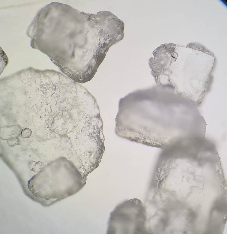
Salt Water Versus Bacteria Under The Microscope

Salt Water Versus Bacteria Under The Microscope Youtube To properly identify bacteria under a microscope, it is essential to first adjust the focus. start by placing the slide on the microscope stage and securing it in place. look through the eyepiece and adjust the focus knob until the image comes into view. move the focus knob slowly to sharpen the image. Once the bacteria are near enough it uses the cilia to push these organisms, along with some water, into the vestibulum. they then move along the buccal cavity until it reaches the mouth (cytostome). from there the bacteria will be acidified and killed. this will make it easier for the bacteria to be digested by the lysosomal enzymes.

Salt Water Versus Bacteria Under The Microscope Youtube Water will flow out of the elodea cells by osmosis, shrinking the cell membrane away from the stiff cell wall (plasmolysis). get a microscope slide. place 2 drops of di water on the left and 2 drops 20% salt on the right. obtain a leaf from a stalk of elodea and cut the leaf in half. place a half leaf in each solution. Some of the other types of bacteria based on morphology include: spirochetes (flexible spiral bacterial) e.g. spirochaetales – spirochetes have an axial filament that allow them to move in a type of motion that has been described as “corkscrew motion”. actinomycetes (branched) e.g. streptosporangineae. Step 2: prepare a neat slide. once the bacterial incubation period is achieved, it is time to prepare a bacterial smear on a glass slide. your slide can be viewed under a microscope to unveil the mysteries of bacteria right after this smear is ready. Escherichia coli. escherichia coli or e.coli is a gram negative species of bacillus shaped bacteria that can be easily observed under a microscope, even for those with the untrained eye. this bacteria has a fast growth rate, doubling every 20 minutes, making them a common choice for bacterial related research purposes.

Salt Under The Microscope вђ Clounce Step 2: prepare a neat slide. once the bacterial incubation period is achieved, it is time to prepare a bacterial smear on a glass slide. your slide can be viewed under a microscope to unveil the mysteries of bacteria right after this smear is ready. Escherichia coli. escherichia coli or e.coli is a gram negative species of bacillus shaped bacteria that can be easily observed under a microscope, even for those with the untrained eye. this bacteria has a fast growth rate, doubling every 20 minutes, making them a common choice for bacterial related research purposes. Place your prepared slides next to each other on a paper towel or staining rack. cover the slides in the chosen stain and leave for 1 2 minutes. remove excess stain by running a gentle stream of water over the surface of the glass slide. place the glass slide on the microscope stage and begin viewing the specimen. Sometimes the liquid used is simply water, but often stains are added to enhance contrast. once the liquid has been added to the slide, a coverslip is placed on top and the specimen is ready for examination under the microscope. the second method of preparing specimens for light microscopy is fixation. the “fixing” of a sample refers to the.

Comments are closed.