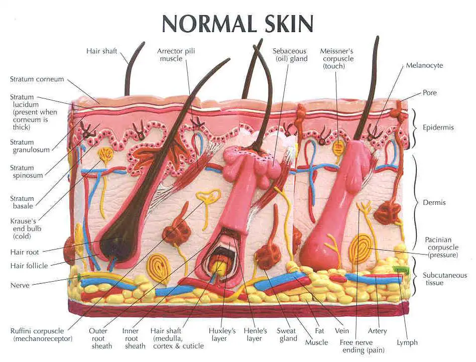
Skin Diagram With Labels

Diagram Of Human Skin Structure вђ Science Learning Hub Skin diagram. the largest organ in the human body is the skin, covering a total area of about 1.8 square meters. the skin is tasked with protecting our body from external elements as well as microbes. the skin is also responsible for maintaining our body temperature – this was apparent in victims who were subjected to the medieval torture of. “thick skin” is found only on the palms of the hands and the soles of the feet. it has a fifth layer, called the stratum lucidum, located between the stratum corneum and the stratum granulosum (figure 5.1.2). figure 5.1.2 – thin skin versus thick skin: these slides show cross sections of the epidermis and dermis of (a) thin and (b) thick.

Skin Anatomy Diagram Labeled Figure 5.2 layers of skin the skin is composed of two main layers: the epidermis, made of closely packed epithelial cells, and the dermis, made of dense, irregular connective tissue that houses blood vessels, hair follicles, sweat glands, and other structures. beneath the dermis lies the hypodermis, which is composed mainly of loose connective. The skin has three basic layers, each with a different role. the number of skin layers that exists depends on how you count them. you have three main layers of skin—the epidermis, dermis, and hypodermis (subcutaneous tissue). within these layers are additional layers. if you count the layers within the layers, the skin has eight or even 10. The skin is composed of two main layers: the epidermis, made of closely packed epithelial cells, and the dermis, made of dense, irregular connective tissue that houses blood vessels, hair follicles, sweat glands, and other structures. beneath the dermis lies the hypodermis, which is composed mainly of loose connective and fatty tissues. This osmosis high yield note provides an overview of skin structures essentials. all osmosis notes are clearly laid out and contain striking images, tables, and diagrams to help visual learners understand complex topics quickly and efficiently. find more information about skin structures: company. about us careers press editorial board blog.

Skin Diagram Labeled The skin is composed of two main layers: the epidermis, made of closely packed epithelial cells, and the dermis, made of dense, irregular connective tissue that houses blood vessels, hair follicles, sweat glands, and other structures. beneath the dermis lies the hypodermis, which is composed mainly of loose connective and fatty tissues. This osmosis high yield note provides an overview of skin structures essentials. all osmosis notes are clearly laid out and contain striking images, tables, and diagrams to help visual learners understand complex topics quickly and efficiently. find more information about skin structures: company. about us careers press editorial board blog. The layers of your skin. your skin includes three layers known as epidermis, dermis, and fat. some health issues, such as dermatitis and infections, can affect how these different layers work to. This video tutorial demonstrates how to draw and label the parts of skin step by step. you can learn how to draw skin diagram easily, just watching this vide.

Comments are closed.