Snapgene Confocal Microscopy Mastersolfe

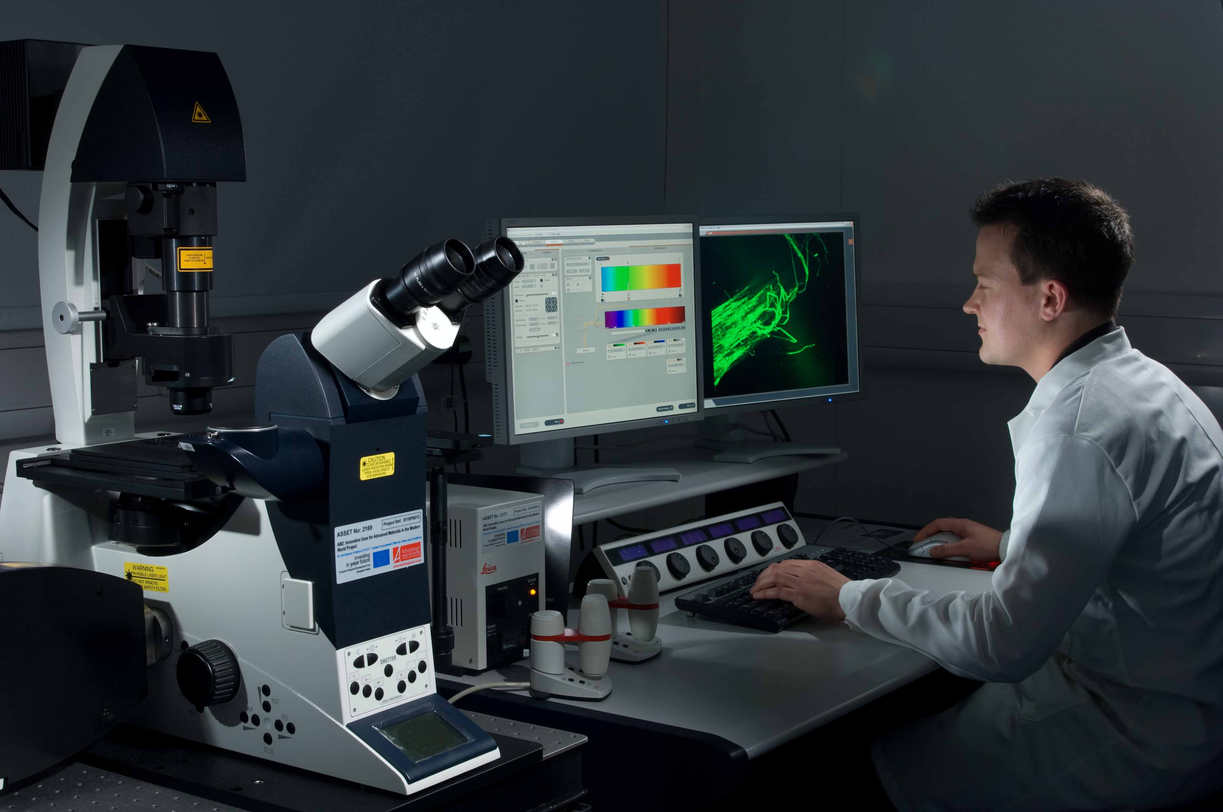
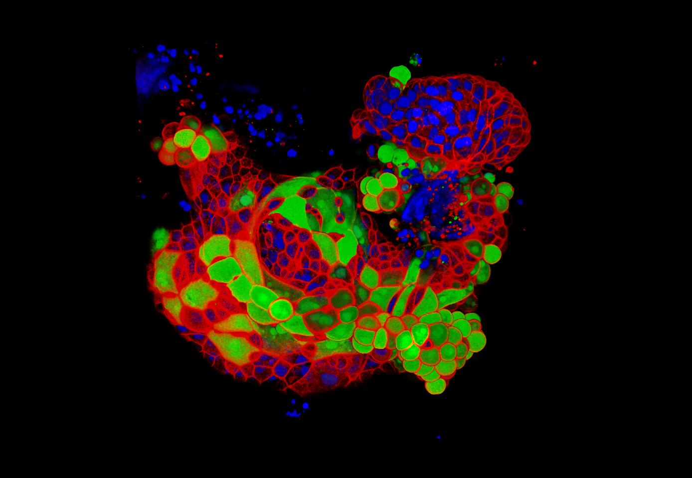
Snapgene Confocal Microscopy Mastersolfe Metrics. when used appropriately, a confocal fluorescence microscope is an excellent tool for making quantitative measurements in cells and tissues. the confocal microscope’s ability to block. The re scan confocal microscope (rcm) is a recently commercialized confocal technology that improves lateral resolution by 1.4 (√2). the rcm includes a re scanning unit consisting of a pair of re scanning mirrors between the pinhole and detector that allows for de coupling of the magnification of the object and scanning spot ( de luca, et al, 2013 ).

Snapgene Confocal Microscopy Mastersolfe The laser scanning confocal microscope (lscm) is an essential tool for many biomedical imaging appli cations at the level of the light microscope. the basic principles of confocal microscopy and the evolution of the lscm into today’s sophisticated instruments are outlined. the major imaging modes of the lscm are introduced including single. The prototype of the lsm 44 series is now on display in the deutsches museum in munich. the lsm 10 – a confocal system with two fluorescence channels. the lsm 310 combines confocal laser scanning microscopy with state of the art computer technology. the lsm 410 is the first inverted micro scope of the lsm family. In this 35 page white paper, learn about the basic principles of confocal microscopy and find a summary of all aspects related to the special nature of image formation in a confocal microscope. download white paper. principle of confocal fluorescence microscopy. optical conditions and the basics of image formation. Laser scanning confocal microscopy serves as a critical instrument for microscopic research in biology. however, it suffers from low imaging speed and high phototoxicity. here we build a novel.

Snapgene Confocal Microscopy Mastersolfe In this 35 page white paper, learn about the basic principles of confocal microscopy and find a summary of all aspects related to the special nature of image formation in a confocal microscope. download white paper. principle of confocal fluorescence microscopy. optical conditions and the basics of image formation. Laser scanning confocal microscopy serves as a critical instrument for microscopic research in biology. however, it suffers from low imaging speed and high phototoxicity. here we build a novel. Intro to confocal microscopy (matt renshaw) auditorium 1 @10:00. intro to live cell imaging (deborah aubyn) seminar room 4 @10:00. intro to optical sectioning (donald bell) seminar room 3 & 4 @11:00. intro to light sheet microscopy (alessandro ciccarelli) seminar room 4 & 5 @10:00. intro to multiphoton microscopy (rocco d’antuono). 2.1 marvin minsky’s microscope. the development of confocal microscopes was, and continues to be, driven by the desire to image biological events as they occur in vivo. the invention of the confocal microscope is attributed to marvin minsky who built a working scanning optical microscope in 1955 with the goal of imaging neural networks in unstained preparations of living brai.

Snapgene Confocal Microscopy Steelnored Intro to confocal microscopy (matt renshaw) auditorium 1 @10:00. intro to live cell imaging (deborah aubyn) seminar room 4 @10:00. intro to optical sectioning (donald bell) seminar room 3 & 4 @11:00. intro to light sheet microscopy (alessandro ciccarelli) seminar room 4 & 5 @10:00. intro to multiphoton microscopy (rocco d’antuono). 2.1 marvin minsky’s microscope. the development of confocal microscopes was, and continues to be, driven by the desire to image biological events as they occur in vivo. the invention of the confocal microscope is attributed to marvin minsky who built a working scanning optical microscope in 1955 with the goal of imaging neural networks in unstained preparations of living brai.

Comments are closed.