The Anatomy Of The Heart Its Structures And Functions

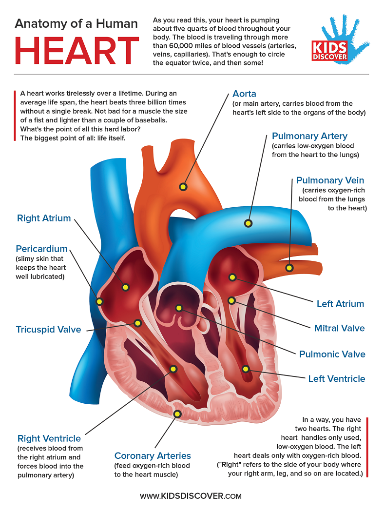
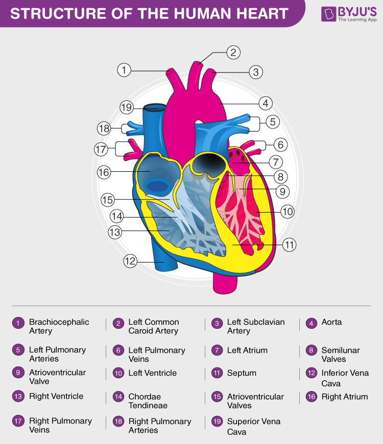
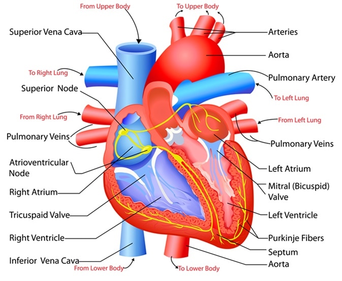
Infographic Anatomy Of The Human Heart Kids Discover The anatomy of the heart, its structures, and functions. the structure of the heart helps supply blood and oxygen to all parts of the body. it is divided by a partition (or septum) into two halves. the halves are, in turn, divided into four chambers. the heart is situated within the chest cavity and surrounded by a fluid filled sac called the. Muscle and tissue make up this powerhouse organ. your heart contains four muscular sections (chambers) that briefly hold blood before moving it. electrical impulses make your heart beat, moving blood through these chambers. your brain and nervous system direct your heart’s function. advertisement.

Human Heart Anatomy Functions And Facts About Heart The aortic semilunar valve is between the left ventricle and the opening of the aorta. it has three semilunar cusps leaflets: left left coronary, right right coronary, and posterior non coronary. in clinical practice, the heart valves can be auscultated, usually by using a stethoscope. Heart, organ that serves as a pump to circulate the blood. it may be a straight tube, as in spiders and annelid worms, or a somewhat more elaborate structure with one or more receiving chambers (atria) and a main pumping chamber (ventricle), as in mollusks. in fishes the heart is a folded tube, with three or four enlarged areas that correspond. The heart is a muscular organ that pumps blood throughout the body. it is located in the middle cavity of the chest, between the lungs. in most people, the heart is located on the left side of the chest, beneath the breastbone. the heart is composed of smooth muscle. it has four chambers which contract in a specific order, allowing the human. Heart anatomy in basic terms. the heart is a crucial organ that functions as the body's pump, ensuring blood circulation throughout the body. it consists of four main chambers: left and right atria (upper chambers) left and right ventricles (lower chambers) these chambers work in a coordinated manner to receive oxygen poor blood, pump it to the.

Structure And Function Of The Heart The heart is a muscular organ that pumps blood throughout the body. it is located in the middle cavity of the chest, between the lungs. in most people, the heart is located on the left side of the chest, beneath the breastbone. the heart is composed of smooth muscle. it has four chambers which contract in a specific order, allowing the human. Heart anatomy in basic terms. the heart is a crucial organ that functions as the body's pump, ensuring blood circulation throughout the body. it consists of four main chambers: left and right atria (upper chambers) left and right ventricles (lower chambers) these chambers work in a coordinated manner to receive oxygen poor blood, pump it to the. Anatomy of the interior of the heart. this image shows the four chambers of the heart and the direction that blood flows through the heart. oxygen poor blood, shown in blue purple, flows into the heart and is pumped out to the lungs. then oxygen rich blood, shown in red, is pumped out to the rest of the body, with the help of the heart valves. The heart is located in the thoracic cavity medial to the lungs and posterior to the sternum. on its superior end, the base of the heart is attached to the aorta, pulmonary arteries and veins, and the vena cava. the inferior tip of the heart, known as the apex, rests just superior to the diaphragm.

Cardiovascular System Anatomy And Physiology Study Guide For Nurses Anatomy of the interior of the heart. this image shows the four chambers of the heart and the direction that blood flows through the heart. oxygen poor blood, shown in blue purple, flows into the heart and is pumped out to the lungs. then oxygen rich blood, shown in red, is pumped out to the rest of the body, with the help of the heart valves. The heart is located in the thoracic cavity medial to the lungs and posterior to the sternum. on its superior end, the base of the heart is attached to the aorta, pulmonary arteries and veins, and the vena cava. the inferior tip of the heart, known as the apex, rests just superior to the diaphragm.

Comments are closed.