The Inferior Mesenteric Artery Position Branches Teachmeanatomy

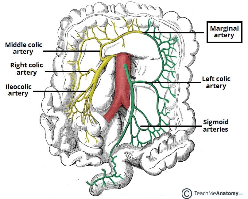
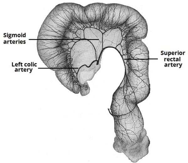
The Inferior Mesenteric Artery Position Branches Teachmeanatomy The branches of the inferior mesenteric artery supply the structures of the embryonic hindgut. these include the distal 1 3 of the transverse colon, splenic flexure, descending colon, sigmoid colon and rectum. there are three major branches that arise from the ima – the left colic artery, sigmoid artery and superior rectal artery. Inferior mesenteric artery – supplies the organs of the hindgut – the distal one third of the transverse colon, splenic flexure, descending colon, sigmoid colon and rectum. the venous drainage of the mesentery is via the superior mesenteric vein (smv) and inferior mesenteric vein (imv), which both run alongside their associated arteries.

The Inferior Mesenteric Artery Position Branches Teachmeanatomy Anatomical position. the superior mesenteric artery is the second of the three major anterior branches of the abdominal aorta (the other two are the coeliac trunk and inferior mesenteric artery). it arises anteriorly from the abdominal aorta at the level of the l1 vertebrae, immediately inferior to the origin of the coeliac trunk. 1 4. synonyms: ima, arteria mesenterica caudalis. the inferior mesenteric artery arises from the abdominal aorta at the level of the third lumbar vertebra. it supplies the hindgut and has four major branches called left colic, sigmoid and superior rectal arteries. it also contributes to the formation of the marginal artery of drummond. Anatomical terminology. [edit on wikidata] in human anatomy, the inferior mesenteric artery (ima) is the third main branch of the abdominal aorta and arises at the level of l3, supplying the large intestine from the distal transverse colon to the upper part of the anal canal. the regions supplied by the ima are the descending colon, the sigmoid. The ima arises from the aorta at the l3 level and has three main branches: the left colic, sigmoid and superior rectal. some variability has been reported in the origin of the three branches. the left colic and sigmoid artery arise independently in 41.2% of patients and share a common trunk in 44.7% of patients.

Comments are closed.