Ultrasound Appearance Of The Superior Mesenteric Artery Abdominal

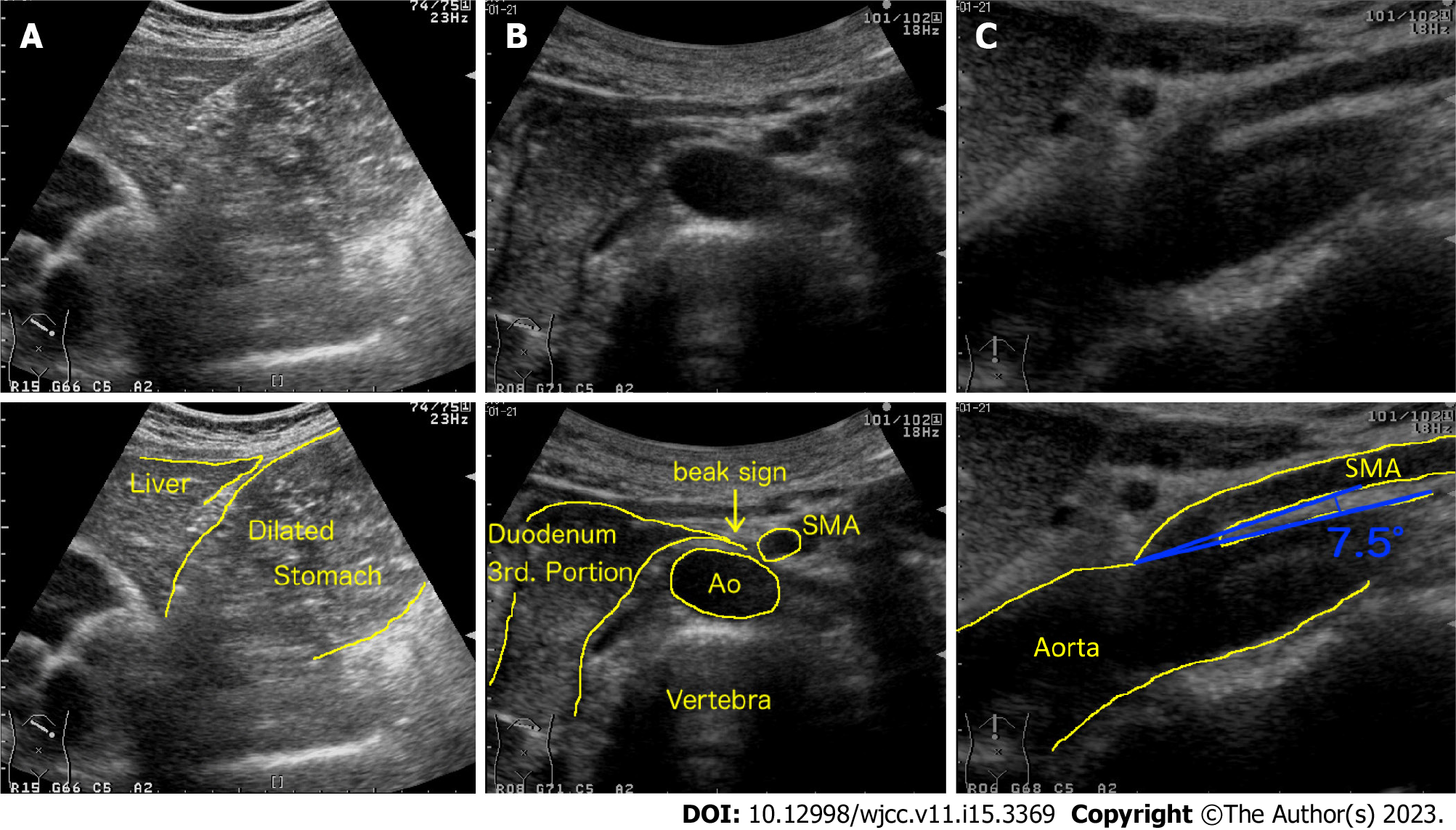
Ultrasound Appearance Of The Superior Mesenteric Artery Abdominal Master course on aortic aneurysm ultrasound abcvascular courses abdominal aortic aneurysm aaa 4 this short video shows the ultrasound appearance. Introduction. superior mesenteric artery (sma) syndrome (or aorto mesenteric compass syndrome or wilkie's disease) [] is a relatively rare condition caused by a short treitz's ligament, or by an unusually low origin of the sma causing a reduction of the angle formed by the aorta and the sma [].

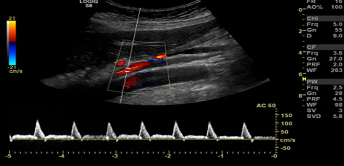
Superior Mesenteric Artery Syndrome Diagnosis And Management Color and pulsed doppler evaluation of the mesenteric arteries is performed to assess for compromise of intestinal blood flow in patients presenting with chronic, unexplained, and atypical abdominal pain. this examination includes evaluation of the abdominal aorta and the celiac, superior mesenteric (sma), and inferior mesenteric (ima) arteries. Ern will be noted of low resistance form.in a normal or mildly obstructed (<50%) sma, peak systolic velocities range from 80 200 cm s, and. nd diastolic flow velocity is < 45 cm s.occlusion of the sma is diagnosed by ultrasound when blood flow is absent in a portion of the vessel du. The superior mesenteric artery (sma) is most commonly affected, likely secondary to its high flow rate and narrow angle of takeoff from the abdominal aorta. most emboli lodge anywhere from 3 to 10 cm distal to the vessel’s origin, typically beyond the branch point of the middle colic artery, which results in sparing of the duodenum and. The superior mesenteric artery is the artery to the midgut. it supplies the gut from the ampulla of vater of the 2 nd part of the duodenum to the distal third of the transverse colon, and includes structures in between such as 5: the inferior pancreaticoduodenal artery also supplies the head of the pancreas.

Superior Mesenteric Artery Ultrasound The superior mesenteric artery (sma) is most commonly affected, likely secondary to its high flow rate and narrow angle of takeoff from the abdominal aorta. most emboli lodge anywhere from 3 to 10 cm distal to the vessel’s origin, typically beyond the branch point of the middle colic artery, which results in sparing of the duodenum and. The superior mesenteric artery is the artery to the midgut. it supplies the gut from the ampulla of vater of the 2 nd part of the duodenum to the distal third of the transverse colon, and includes structures in between such as 5: the inferior pancreaticoduodenal artery also supplies the head of the pancreas. An aortomesenteric angle (between the superior mesenteric artery [sma] and aorta; fig. 1c) of less than 41° is 100% sensitive and 55.6% specific for nutcracker syndrome; a normal aortomesenteric angle measures approximately 90° . a beak angle measurement may also be obtained but is cumbersome to perform. The superior mesenteric artery (sma) originates approximately 1.5–2 cm below the celiac artery, crossing anterior to the third segment of the duodenum and subsequently entering the mesenteric root before giving rise to 12–16 jejunal and ileal branches. the sma usually measures about 6–8 mm in anteroposterior diameter in its proximal segment.

Comments are closed.